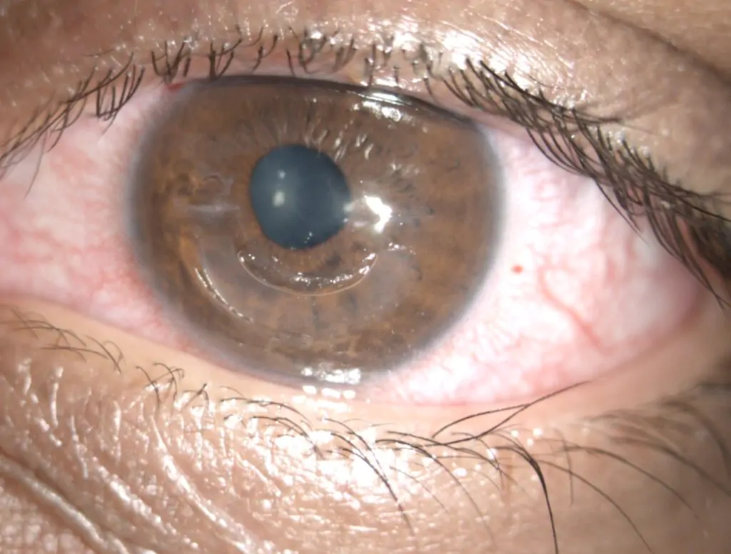
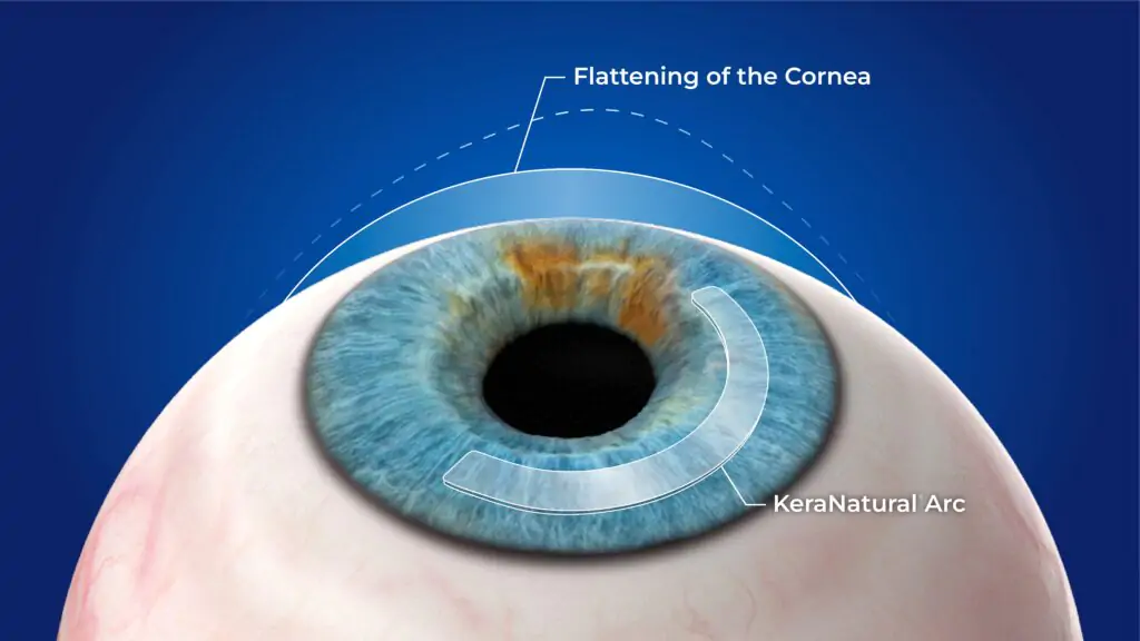
Keratoconus (KC) is a progressive corneal ectasia and weakening with onset typically in the second decade of life. As keratoconus progresses, irreversible visual loss occurs as the result of increasing irregular corneal astigmatism, and the quality of life declines in patients.
Keratoconus is a common corneal disease and it is more prevalent in the area of West India and the Middle East which indicate a possible genetic predisposing for this condition.
The corneal stroma accounts for approximately 80% of the corneal thickness and it is usually made of highly organised collagen layers,
which contribute to the clarity of this live tissue. Recent evidence showed increased inflammatory (granular) cells in KC corneas and they may play a role in the breakdown and phagocytosis of corneal tissue, which support the inflammatory theory as a contributor to keratoconus progression. Rarely, corneal ectasia could also develop following laser refractive surgery, where the strength of the cornea has been weakened, which allows the cornea’s shape to change and become irregular.
Mild cases of KC could be treated with spectacles and observation, or collagen cross linking to stop the progression of the condition. When spectacles are not enough to improve vision, the next step is contact lenses, however not all the patients are suitable for contact lenses and they are not without side effects like causing corneal infection and scarring.


