Corneal And Anterior Segment Conditions & Treatment

Cataracts

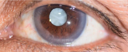
A cataract is the clouding of the lens inside the eye. The most common cause is aging, and by the age of 60 year, almost half of the population develop some form of cataract. The key symptom is blurred vision that worsens over a period of months or years. Some people also complain of glare under sun light or when facing car headlights when driving in the evening. Colours become less distinct. We provide cataract surgery to clear the vision. Also we can provide solution for patients who would like to be independent from glasses with touching up with Laser refractive procedure (Presbyond procedure)

Corneal pathology

Cornea is the clear window of the eye that covers the iris. It is considered the most powerful lens in the eye. The cornea is kept moist by the tear film and any pathology that would affect the clarity or the shape of the cornea would result of blurred vision. Dry eyes are the most encountered pathology of the eye, and it usually present with the sensation of irritation and grittiness and blurred vision. Dry eyes could be related to low tear production or to eye exposure, which is mostly due to lid malposition. Various procedures could be applied to reduce the exposure of the eye, with surgical correction of lid malposition or sometimes with fillers.

Corneal Dystrophies

Are a variety of hereditary corneal diseases that affect corneal clarity and hence the vision. Corneal dystrophy could affect any part of the cornea and the most famous corneal dystrophy is the Fuch’s dystrophy, which affects the endothelium layer, the most posterior part of cornea and is considered the pumping machine of the cornea that maintain the clarity of the cornea.

Corneal ectatic disorders

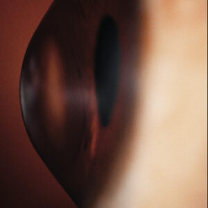
The usual corneal shape is almost spherical, however in case of ecstatic disorder, the cornea becomes thinner with protrusion and distortion of the anterior corneal surface, hence disturbance of vision. Depending on the shape the cornea assumes in ectatic disorder, the cornea may be shaped as a cone (Keratoconus), as a large thin cornea (Keratoglobus), or have a peripheral thinning (Pellucid marginal corneal degeneration).